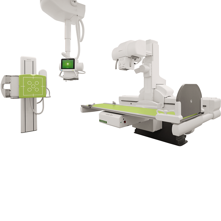
| 1 | Table | 1 | ||
| Dimensions Table height Elevation range Elevation speed Movements Tilt angle Tilt speed Automatic stop |
: ±62 cm – 142 cm (24.4″ – 59.9″), elevating, motorized : ±80 cm (31.5″) : 2.5 cm/s (1″/s) : – 90° to + 90° : 2 speeds, 4.5°/s and 6.5°/s : In horizontal position |
|||
| 2 Table top | 1 | |||
| Dimensions | ||||
| Total dimensions | : ±73.8 cm x 235.6 cm (29″ x 92.8″) | |||
| Radiolucent area | : ±221.4 cm x 55.4 cm (87.2″ x 21.8″) | |||
| Table top to detector distance | : Min. 10.2 cm (4″) | |||
| Material | : Micro sandwich of laminate, carbon and foam | |||
| X-ray attenuation | : 0.6 mm (0.02″) Al (at 100 kVp, HVL = 2.7 mm (0.1″) Al) | |||
| Maximum patient weight without limitations in movements | : ±284 kg (626 lbs) | |||
| Movements | ||||
| Lateral | : ± 32 cm (12.6″) = (± 16 cm (6.3″)) | |||
| Longitudinal | : Only detector movements to improve patient comfort | |||
| Movement speed | ||||
| Lateral | : ±5 cm/s (1.9″/s), soft start and stop Auto centering | |||
| Longitudinal | : Detector movement 3 cm/s – 20 cm/s (1.2″/s – 7.9″/s) | |||
| Tube Column-detector Assembly | ||||
| Movements | ||||
| Range | : ±160 cm (63″) longitudinal, motorized | |||
| Speed | : Variable, slow for positioning, high for travel maximum speed from 3 cm – 20 cm/s (1.2″/s – 7.9″/s) | |||
| Patient coverage | : ±203 cm (79.9″), without patient movement | |||
| Angulation | ||||
| Movements | : Motorized | |||
| Range | : ± 40° | |||
| Speed | : ±11.2°/s | |||
| Supporting functions | : Automatic centering of target organ during oblique projections in fluoroscopy | |||
| : Oblique exposures at both ends of tabletop possible | ||||
| Source image distance | ||||
| Range | : ±113 cm – 183 cm (44″ – 72″) | |||
| Movements | : Motorized | |||
| Speed | : ±4.1 cm/s (1.6″/s) | |||
| Focal spot to floor distance (in 90° position) | : ±51.5 cm – 211.5 cm (20.3″ – 83.3″) (without angulation) | |||
| Tube rotation | : Manual | |||
| Range | : – 90°/ + 180° | |||
| Stop position | : – 90°/ – 50°/ – 40°/ 0°/ + 40°/ + 50°/ + 90°/ + 180° | |||
| 3 | 7 Amplimat chamber on the fluoroscopy table | Yes, this usefull for driving the x-ray beam with AEC for easy positioning patient and guaranty there have adequate: x-ray dose for better image quality | 1 | |
| 4 | Compressor | 1 | ||
| Movements | : | Motorized | ||
| Activation | : | Remote controlled | ||
| Compression force | : | Variable, 3 kg – 15 kg (6.6 lbs – 33 lbs) in 0.5 kg (1.1 lbs) steps | ||
| Compressor parking | : | Automatic, behind tube stand | ||
| Cone | : Removable | |||
| 5 | Grid | : Stationary Grid | 2 | |
| Material | : Carbon fiber | |||
| Lines | : 44 lines/cm | |||
| Focus (FO) | : ±120 cm (47.2″) and 180 cm (70.9″) | |||
| Automatic selection of Grid according to SID and programmed examination plus motorized removal of grids. Fully | ||||
| automatic, no grid handling necessary | ||||
| 6 | Single monitor trolley | : the trolley including 21.3″ medical monitor, articulated, height adjustable. | 1 | |
| 7 | Nearby control trolley | Trolley for in-room control of imaging and geometry parameters, including touchscreen geometry control console,: footswitch for fluoroscopy and exposure and sealed waterproof keyboard with touchpad. | 1 | |
| 8 | X-ray generation | 1 | ||
| Exposure output power | : | 65 kW | ||
| kV | : | 40 – 125 kV (dynamic exposures) | ||
| mA range | : | 1 – 650 mA | ||
| ms range | : | 1 ms – 4 s with AEC (Automatic Exposure Control) | ||
| ms range | : 1 ms – 16 s without AEC | |||
| Manual mode | ||||
| Two-factor technique (kV – mAs) | : Yes | |||
| Three-factor technique (kV – mA – s) | : Yes | |||
| Automatic mode | ||||
| One-factor falling load (kV) | : Yes | |||
| Two-factor constant load (kV/mA) | : Yes | |||
| Automatic kV reduction | : Yes | |||
| IQX provides excellent, reliable and consistent image quality for digital exposures, both in static and dynamic | ||||
| fluoroscopy studies. IQX controls and adapts the exposure parameters within the X-ray pulse. The automatic and | ||||
| IQX Intelligent exposur | : | fast regulation of kV during each exposure leads to high image quality for all types of studies, for all patients. | ||
| Control Fluoroscopy | : Yes | |||
| 9 | Tubes on Fluoroscopy table | : | 1 | |
| Focal spot (small / large) | : 0.6 / 1.2 | |||
| Min. anode speed | : up to 10K revolutions/minute | |||
| Anode angle | : ±13° | |||
| Maximum voltage | : | ±125 kV or (110 kV with GCF) | |
| Nominal radiographic anode input power | : | ±30 kW / 85 kW | |
| Double tube overload protection | : | Yes | |
| Total filtration minimum | : | 2.5 mm AI (IEC 60522, 75 kV) | |
| Compatible with pulsed fluoroscopy | : | Yes | |
| Compatible with Controlled Fluoroscopy | : Yes | ||
| 10 Detectors (Fix on Fluoroscopy table) | 1 | ||
| Electronic flat detector (CsI/Cesium Iodide) | : | Yes | |
| Maximum field of view | : | ±43 cm x 43 cm (17″ x 17″) | |
| Image matrix | : | ±2880 x 2881 pixels | |
| Pixel size | : | 148 μm | |
| Bit depth | : | 16 bits | |
| DQE | : | 65 % at 0.1 lp/mm | |
| MTF | : | 55 % at 1 lp/mm | |
| Exposure dose | : | 0.1 – 10 μGy | |
| Cooling | : | Automatic internal air cooling system | |
| 11 Automatic Image stitching on fluoroscopy table | : Yes | 1 | |
| Capability doing Automatic image stitching, no need additional bucky stand but can using fluoroscopy table we call | |||
| paralel stitching. As for standing (table tilt to standing 90 ° or patient or stitching in vertical posistion also laying | |||
| : | down on the fluoroscopy table and the stitching in horizontal projection) | ||
| 12 Eleva workspot | 1 | ||
| Processor Computer | : Based on Intel Core I7 | ||
| Memory | : 16 GB RAM | ||
| Local storage | : 1 TB Solid State Disk (SSD) | ||
| Image review | |||
| Dynamic unique for fluoroscopy and radiography | : Yes | ||
| Multi image review | : Mosaic view of 4, 9 or 16 single images | ||
| Zoom 1:1, 2:1, 3:3 | : Yes | ||
| Monitors | |||
| Two high quality color LCD monitors, one with touchscreen | : | Yes | |
| Size | : | 21.3″ | |
| DICOM calibrated for room environmental illuminance from 0 to 1000 LUX | : | Yes | |
| DICOM illuminance compensation automatically adjusted for room illuminance | : | Yes | |
| 13 UNIQUE2 Dynamic UNIQUE | : Yes | 1 | |
| 2nd generation of post processing software that automatically adjust contras and enhance image detail, optiming | |||
| image processing that reducing motion blur (leg effec) and optimaling image from the first frame so no initial dose | |||
| needed. Adjust contras and minimizing noise. | |||
| 14 DICOM | 1 | ||
| DICOM interoperability | : | Yes | |
| DICOM Print for Rad images | : | Yes | |
| DICOM store DICOM storage commit | : | Yes | |
| DICOM worklist management | : | Yes | |
| DICOM MPPS | : Yes | ||
| 15 Monitor to be placed in examination room | 1 | ||
| Type | : | LCD color monitor IPS | |
| Size | : | 21.3″ | |
| Matrix | : | 1600″ x 1200″ (2 Megapixel) | |
| Luminance ratio | : | > 800:1 | |
| Dimensions | : | approx. 495 x 425 mm (19.5″ x 16.7″) | |
| Weight | : | approx. 7.1 kg (15.6 lbs) | |
| DICOM calibrated for room environmental illuminance from 0 to 1000 LUX | : | Yes | |
| DICOM illuminance compensation automatically adjusted for room illuminance | : Yes | ||
| Acessories | |||
| 16 Handgrips | : | Yes | 1 |
| 17 Shoulder support | : | Yes | 1 |
| 18 Side bar | : | Yes | 1 |
| 19 Pair of leg supports | : | Yes | 1 |
| 20 Dry Printer | : | Yes | 1 |
| 21 Panel & Cable | : | Yes | 1 |
| 22 UPS 3kVA | : | Yes | |
| 23 Warranty (year) | : Yes | 2 | |


