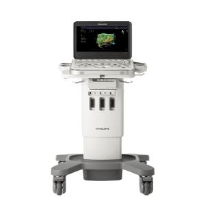
| SPECIFICATION Philips EPIQ Elite Diagnostic Ultrasound System GI Basic | ||
| ULTRASONOGRAPHY SYSTEM | ||
| I. GENERAL INFORMATION | ||
| SOLE AGENT/DISTRIBUTOR | PT PHILIPS INDONESIA COMMERCIAL | |
| ADDRESS | Cibis Nine 10 th Floor. Jl. T.B. Simatupang No.2, Jakarta 12560 | |
| PHONE/ FAX. / E-MAIL | 021- 29651333 | |
| NAME OF PRODUCT | Philips EPIQ Elite Diagnostic Ultrasound System GI Basic | |
| BRAND/MANUFACTURER | Philips | |
| MODEL/TYPE | EPIQ Elite | |
| COUNTRY OF ORIGIN | USA | |
| FACTORY OF ORIGIN | USA | |
| PRODUCT’S REGISTRATION (AKL) | AKL 21501024221 | |
| II. | MAIN COMPOSITION | Main Unit with 21,5inch wide format high definition 2 customize probe (3D9-3v / C5-1 / C8-5 / C9-2 / C10-3v / C10-4ec / D2cwc / D2tcd / D5cwc / eL18- 4 / L12-3 / L12-3 ERGO / L12-5 50 MM / L12-5 / L18-5 / mC7-2 / mC12-3 / S5-1 / S8-3 / S9-2 / S12-4 / V9-2) IMT MFI GI3DQ Battery Ext Life Package Strain Elastography ElastPQ ElastQ Imaging DICOM MaxVue BW Printer Color Printer UPS 3 KVA |
| III. | STANDARD FEATURES | XRES |
| SonoCT | ||
| THI (Tissue Harmonic Imaging) | ||
| Panoramic 2D | ||
| Cine | ||
| Live Compare | ||
| Tissue Aberration Correction (TAC) | ||
| HighQ (automatic Doppler Analysis) | ||
| iSCAN intelligent scanning | ||
| AutoSCAN (real time frame by frame TGC optimization) | ||
| Auto Flow Tracking | ||
| Console Design | 1 TB hard drive | |
| 8 TGC (Time Gain Compensation) slides | ||
| On-board control panel touch screen | ||
| Unique easy-clip cable management solution | ||
| Up and down 10 in/25.4 cm | ||
| Rotates 180 o from center | ||
| USER INTERFACE | ||
| Monitor | Main Unit with 21,5 inch wide format high definition | |
| Extended viewing angle >178 o (horizontal and vertical) | ||
| Response time <14 ms | ||
| Virtually flicker-free technology that reduces eyestrain | ||
| Mounted on fully articulating extension arm | ||
| Control Panel | Full-color 12-inch capacitive touch screen Complete with swipe technology |
|
| SYSTEM OVERVIEW Clinical Application |
• Abdominal • Vascular • Gynecology • Pediatrics GI • Small Parts • Musculoskeletal • Obstetric • Urology • Interventional |
| Supported Transducer Types | Convex Array Probe |
| Linear Array Probe | |
| Micro Convex Array Probe | |
| Supported Transducer Types (Optionals) | Intraoperative Array Probe |
| Pedoff Array Probe | |
| Volume Convex Array Probe 94D) | |
| Sector Array Probe | |
| Imaging Mode | 2D mode |
| M mode | |
| Color mode | |
| Color Power Angio (CPA) | |
| Pulsed Wave Doppler | |
| Duplex mode (Simultaneous 2D and Doppler) | |
| Triplex mode (Simultaneous 2D, Doppler and Color or CPA) | |
| SYSTEM OPTION Electrical Power |
Voltage: 100 to 240 VA Power consumption < 600 VA Frequency; 50 to 60 Hz |
| Dimension | 146 – 171.5 x 57.2 x 109.2 (H x W x D) Weight 104,3 kg without peripheral devices |


